A spinning disk microscope is a type of advanced microscopy system that uses a spinning disk with multiple pinholes to create an optical section of a sample. This enables high-speed imaging of living cells and tissues in three dimensions with minimal phototoxicity and photobleaching. The spinning disk system consists of a spinning disk with a set of pinholes, a motor to rotate the disk, and an optical system to direct the excitation light to the sample and collect the emitted light. The pinholes create a pattern of illumination that sweeps across the sample, allowing only a thin slice of the sample to be illuminated at any given time. The emitted light from the illuminated slice is then detected by a camera and used to construct a 3D image of the sample. Spinning disk microscopy is particularly useful for studying dynamic biological processes, such as intracellular transport, cell division, and vesicle trafficking.
PHOTIK SCIENTIFIC PVT. LTD.
Technology is the future
Phone
+91-9310249352 / +91-8605504814
+91-9310249352 / +91-8605504814
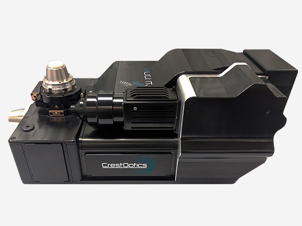
Spinning Disk